
 Yahoo News
 '3D mammography' could transform breast cancer screening and save thousands from unnecessary biopsies
A pioneering “3D mammogram” technique could halve the number of women unnecessarily sent for potentially painful breast biopsies, an NHS study shows.
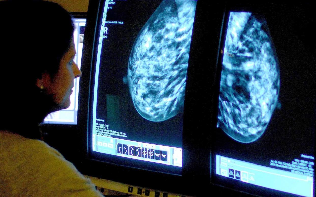
A trial of more than 30,000 women in Leeds found that taking multiple images of the breast from different angles enabled a computer to build accurate 3D images of tissue where cancer was suspected.
If rolled out across the NHS, the technology could mean thousands fewer women have to undergo invasive surgery only for doctors to find that they were not suffering from cancer.
Published in the journal Radiology, the trial at Leeds Teaching Hospital NHS Trust showed that of 827 women two were recalled for further testing after an initial conventional scan indicated an abnormality, 571 required a biopsy.
Of these, 429 - 75 per cent - showed the abnormality was benign.
Blinded to the original results, the researchers then decided whether or not they would have ordered biopsies based on the more accurate 3D images.
The inclusion of the 3D imaging, known as digital breast tomosynthesis, reduced the number of biopsies that would have been ordered by nearly half.
"DBT allows for improved reader accuracy and confidence in determining if a mammographic abnormality is concerning or not, leading to a reduction in the number of biopsies performed," said Dr Nisha Sharma, who led the research.
“Our study validates that DBT can help in the diagnostic workup of mammographic abnormalities and reduce harm to women through fewer false positive biopsies without any reduction in the cancer detection rate."

