
 Yahoo News
 Surgeons trial 'glowing' fluorescent marker to help spot dangerous brain tumour cells

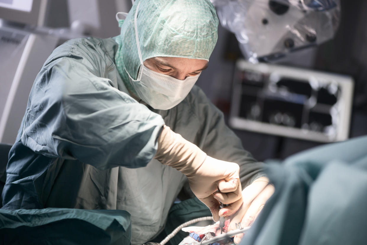
A ‘fluorescent’ chemical that glows pink when a light is shone on it could help surgeons remove dangerous brain tumour cells more accurately.
Surgeons have trialled the fluorescent market, called 5-aminolevulinic acid or 5-ALA, to see if it can help show cancerous cells in the brain so they can remove them.
The research was carried out with patients who had suspected glioblastoma, the most common form of brain cancer.
Treatment usually involves surgery to remove as much of the cancer as possible, but it can be difficult for surgeons to identify all of the cancer cells while avoiding healthy brain tissue.
Previous research shows that, when consumed, 5-ALA accumulates in fast growing cancer cells and this means it can act as a fluorescent marker of high-grade cells.
The latest study saw 99 patients with suspected high-grade gliomas given a drink containing 5-ALA.
MORE: Investigation underway after children hospitalised when inflatable slide at fireworks display collapsed
MORE: Paraplegic man ‘forced to drag himself’ through Luton Airport
Surgeons then used operating microscopes to help them look for fluorescent tissue while removing tumours from the patients’ brains.
The tissue they removed was sent to the pathology lab where scientists could confirm the accuracy of the surgeons’ work.

During their operations, surgeons reported seeing fluorescence in 85 patients and 81 of these were subsequently confirmed by pathologists to have high-grade disease. One was found to have low-grade disease and three could not be assessed.
In the 14 patients where surgeons did not see any fluorescence, only seven tumours could be subsequently evaluated by pathology but in all these cases, low-grade disease was confirmed.
Colin Watts, professor of neurosurgery and chairman of the Brain Cancer Programme at the University of Birmingham, who led the study, said: “Neurosurgeons need to be able to distinguish tumour tissue from other brain tissue, especially when the tumour contains fast-growing, high-grade cancer cells.
“The advantage of this technique is that it may highlight more quickly high-grade disease within a tumour during neurosurgery.
“What this means is that more of the tumour can be removed more safely and with fewer complications, and that’s better for the patient.”
The trial is being presented at the 2018 National Cancer Research Institute (NCRI) Cancer Conference in Glasgow.

